Research
I ask what makes neurons and circuits vulnerable or resilient when they are pushed beyond their normal operating range, whether by energy failure in stroke or by neurodegeneration in Parkinson's disease.
My models are biophysically detailed and multiscale: they include ion channels, pumps and exchangers, the diffusion of ions and metabolites inside cells and through the extracellular space, oxygen supply and cellular metabolism. Building them relies on the reaction-diffusion module of the NEURON simulator, which I helped develop (see Software).
Spreading depolarization across scales
A mismatch between energy supply and demand can trigger spreading depolarization (SD), a slowly propagating wave of near-complete depolarization that occurs in ischemic stroke, migraine aura and traumatic brain injury.
Dendrites
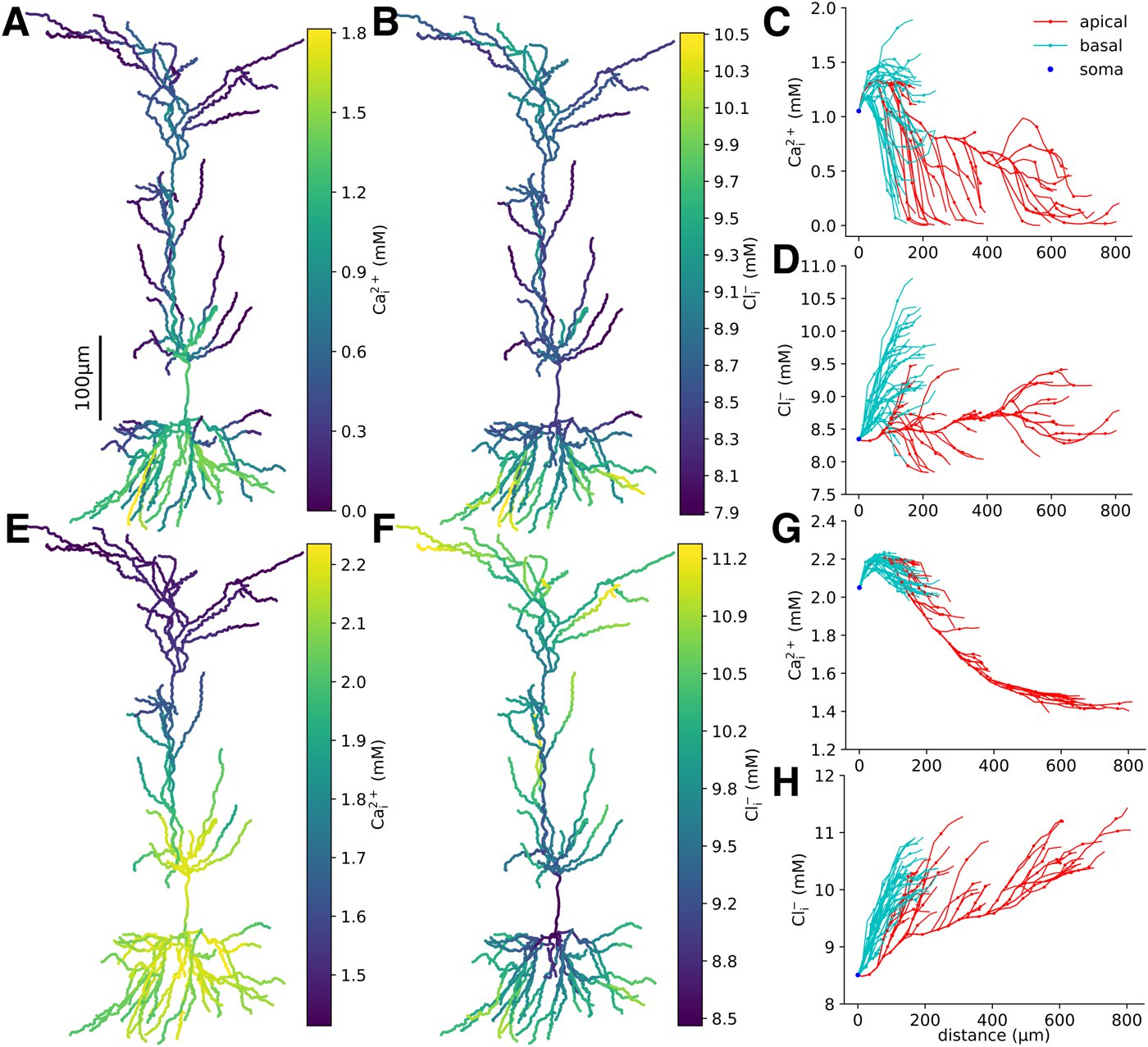
In a morphologically detailed model of a hippocampal CA1 pyramidal neuron, I showed that dendritic compartments experience ischemia and SD differently. Calcium accumulated most in the basal dendrites under both conditions, suggesting they are most exposed to excitotoxicity, while in ischemia, but not SD, the distal apical dendrites saw the highest intracellular chloride, which may lead to dendritic beading (Newton et al., 2026, PLOS Comput Biol).
Brain slices
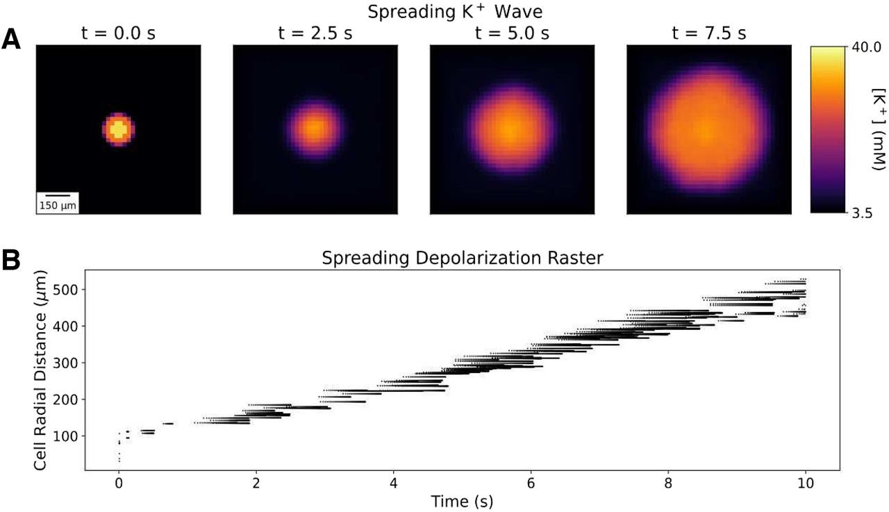
At the tissue scale, a model of 36,000 neurons embedded in the extracellular space of a brain slice linked SD susceptibility to cytoarchitecture, particularly neuronal density and extracellular volume, and showed that neurons near the oxygenated slice surface sustain firing longer (Kelley et al., 2022, eNeuro).
Cortex in vivo
I am now extending the tissue-scale model to SD in vivo: a cortical microcircuit with Potjans-Diesmann connectivity in which neurons and capillaries are placed according to immunohistochemical staining of human cortex (Wang et al., 2026, Microcirculation), each neuron has a detailed model of cellular metabolism, and the Na+/K+-ATPase follows the Post-Albers cycle so that pump rate depends on ion concentrations, membrane potential and available energy.
Cell-to-cell variability in Parkinsonian motor cortex
Pyramidal tract neurons in layer 5B of mouse primary motor cortex are the main cortical output to the brainstem and spinal cord. In Parkinsonian mice they become less excitable, but the change varies widely between neurons. Using recordings from Hong-Yuan Chu's lab, I use data-driven optimization to fit multiple models to each neuron. The models capture both individual responses and the heterogeneity of the population, and they are degenerate: different combinations of conductances reproduce the same behaviour.
Future directions
When is spreading depolarization harmful?
SD contributes to tissue damage after ischemic stroke, but the spreading depression that follows can be benign or even protective. I will extend my models from the initial phase of SD to the NMDA-receptor-driven late phase and the silent period that follows, including neurite beading, Zn2+ handling, K2P leak channels and astrocytic K+ buffering, to identify the conditions that decide which outcome occurs and point to neuroprotective strategies.
How does blood flow turn SD into spreading ischemia?
Under physiological conditions depolarization triggers a hyperemic response; in pathological conditions SD can instead cause vasoconstriction and spreading ischemia. I will add two-way coupling between vessels and tissue through competing vasoactive signals so the model captures the switch from normal to inverse neurovascular coupling as a function of metabolic state, and test which interventions prevent it.
Does cellular diversity protect the Parkinsonian circuit?
Comparing models fit to control and Parkinsonian neurons will identify the conductance changes behind the loss of excitability, and whether different neurons reach the same phenotype through different channel changes, which matters for therapies that target a single channel. Placing these heterogeneous populations into a biophysically detailed M1 network will test whether cellular diversity protects against pathological beta synchrony.
Earlier work
- Adenosine and biosensors. For my PhD I modeled adenosine clearance in neural tissue and showed that free-flow calibration of microelectrode biosensors can substantially underestimate tissue concentrations (Newton et al., 2017, J Neurophysiol).
- Synaptic specificity. 3D intracellular reaction-diffusion simulations of PKMζ spread tested conditions for synaptic specificity during the maintenance of plasticity (Huertas et al., 2022, eNeuro).
- Cardiac nervous system. Biophysical models of intrinsic cardiac neurons based on single-cell transcriptomics (Gupta et al., 2025, J Physiol).
- Biomedical text mining. A pipeline to retrieve and extract COVID-19 immune signatures from the research literature (Newton et al., 2023, BMC Bioinformatics).